
胫腓骨骨折
还会造成足趾畸形?

警惕“马缰绳畸形”
近日,一名男性患儿因“胫腓骨骨折术后出现足趾背伸畸形5月余”入院进行步态检查,主诉在行走约100米后感足趾疼痛不适,并且逐渐加重,出现行走困难,严重影响生活。
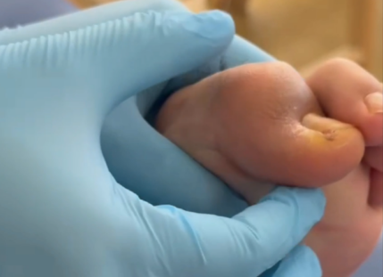
物理评估发现:患儿自然站立位及步行过程中,左侧第一足趾垂状畸形,左侧踝关节被动背屈时,第一足趾屈曲畸形加重。左侧踝关节被动跖屈时,第一足趾屈曲畸形减轻。

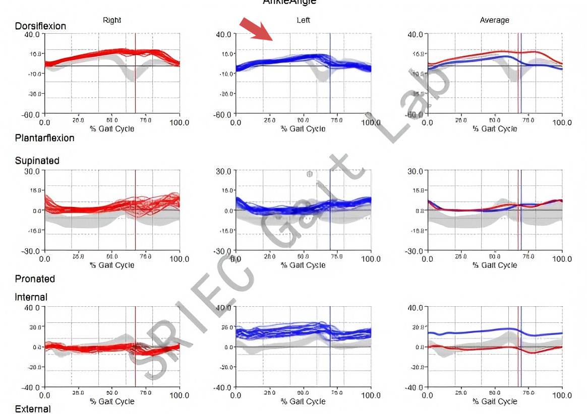
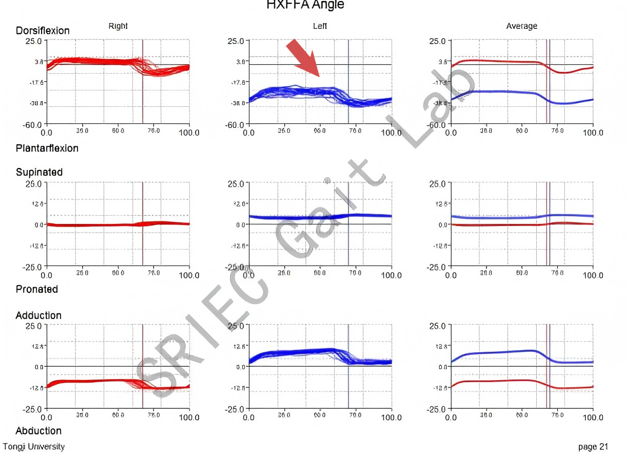
步态分析结果发现:步行过程中,左侧踝关节活动度降低,足大趾活动度降低,为过度屈曲状态,且内收角度异常增加。
PART 01
什么是马缰绳畸形?
马缰绳畸形(checkrein deformity )是一种少见的创伤后遗症,Clawson[1] 在 1974 年最早描述了胫骨骨折后发生的爪形趾,Jahss[2] 在 1992 年将这种常发生于踇长屈肌腱 (flexor hallucis longus,FHL) 及趾长屈肌腱 (flexor digitorum longus,FDL) 的挛缩或“肌腱固定现象”形容为“checkrein deformity ”,即“马缰绳”畸形,其特征性表现为跖趾关节的背伸而趾间关节屈曲,在踝关节背伸时出现爪形趾畸形,而踝关节跖屈时足趾可以伸直,类似骑手骑马时通过马缰绳控制马头,防止马头过低的动作。

PART 02
马缰绳畸形病因
马缰绳畸形通常发生在胫腓骨骨折、踝关节骨折脱位、距骨骨折脱位、跟骨骨折等创伤后或手术后,或亚临床型的小腿后部深筋膜间室综合征。其机制与踇长屈肌腱、趾长屈肌腱的损伤、粘连、挛缩有关[3]
PART 03
解剖学基础
踇长屈肌腱(FHL)与趾长屈肌腱(FDL)
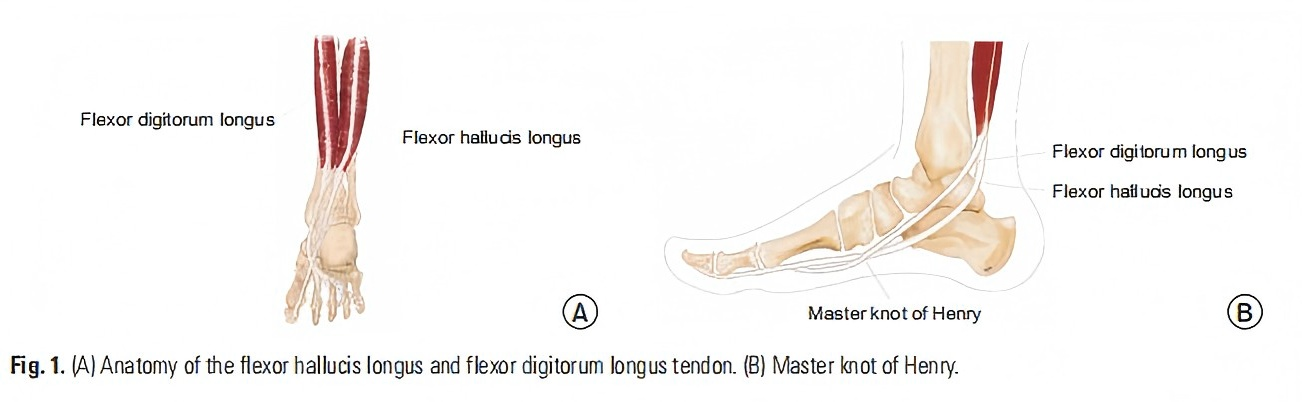
FHL是小腿深部肌肉(图1A)。它起源于腓骨后部远端的一半,沿内侧下方经过胫骨远端后方及内踝后方的沟槽,止于远节趾骨基部[4]。其主要作用是足大趾屈曲。肌腱部分起始于内踝后方上方,位于胫后肌和趾长屈肌腱(FDL)的外侧后方。同时,FDL是小腿深部的一个肌肉,它起源于FHL起点的远端胫骨内侧,在中足处分为四条肌腱,止于第二至第五远节趾骨基部[5]。主要作用是小脚趾屈曲。
FHL能轻松附着于骨折部位的骨痂,特别是胫骨远端或有时腓骨骨折处[6]。此外,由于FHL的肌肉体积大于远端胫骨后的FDL,因此FHL更常受到影响[7]。FHL的肌腱在此处与大脚趾肌腱的附着点之间形成弓弦结构[8]。因此,当肌腱受压时,远端指骨基部弯曲,导致爪形趾畸形。
Henry 结
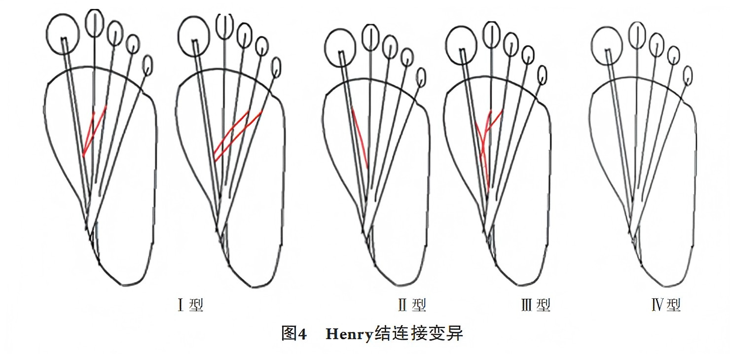
解剖学上,FHL和FDL肌腱在跟骨结节下方交叉的部位,称为“Henry结”。在 Henry 结部位,FHL 与 FDL之间存在腱性连接的变异 (图 4)。 O'Sullivan[9]等认为, 在 Henry 结处FHL与FDL有4种连接的变异。其中连接至第 2、3 趾的相对固定,至第 4、5 趾的也有一定比例;Ⅱ、Ⅲ 型较少;Ⅳ型极少。因此, 单纯的 FHL 粘连不仅会引起趾屈曲畸形 (Ⅱ、Ⅲ 型 ),还可能同时引起其他 4 趾不同程度的屈曲畸形 (I、Ⅲ型)。
PART 04
诊断
病史采集
询问患者是否有可能导致 FHL或 FDL肌腱短缩或卡压的骨折史、室间隔综合征以及跟腱延长术等手术史。
患者可能会抱怨行走时脚趾抓地,脚趾远端疼痛。
踝关节或踝关节外翻会引起踝关节后内侧疼痛,从而导致FDL紧张[10]。
体格检查
踝关节被动背屈踇趾趾间屈曲畸形更加明显,且不能通过被动力伸展。
当踝关节跖屈时,屈曲畸形完全矫正[3]。
鉴别诊断
槌状趾、锤状趾和爪状趾应作为鉴别诊断。
槌状趾:远端趾间关节屈曲,而跖趾关节则不受影响。
锤状趾:近端趾间关节屈曲,跖趾关节中立位或背伸,远侧趾间关节中立位或背伸。
爪形趾:跖趾关节过度伸展的,而近端和远端指间关节则固定在屈曲位置。
PART 05
治疗
保守治疗
对于马缰绳畸形的治疗,一开始可以尝试保守的方法,第一种方法是被动牵伸训练。
另外,患者可使用针对性鞋垫或脚趾支撑器来缓解部分症状[11]。
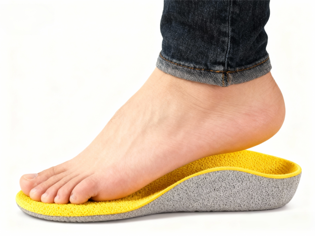
(图片来源于AI,仅供参考)
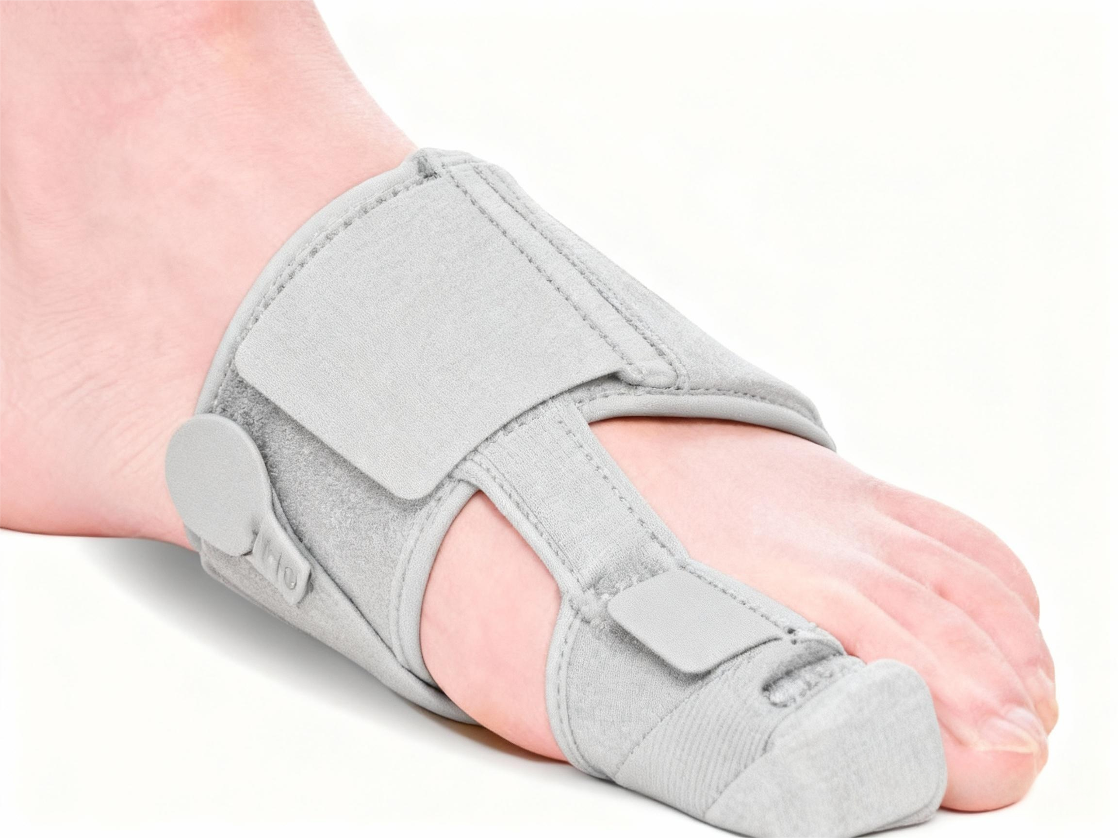
(图片来源于AI,仅供参考)
手术治疗
各种手术选择可用于纠正马缰绳畸形。包括:
骨折部位粘连松解[6,10,12]。
骨折部位Z字成形术延长FHL并松解粘连[13,14]。
中足Z字成形术延长FHL,但不解除粘连[7,15,16]。
踝后或跗管附近Z字成形术延长FHL[11,17,18]以及
FHL肌腱切开术联合指间关节融合术治疗复发病例[19]。
参考文献:
[1] Clawson DK . Claw toes following tibial fracture[ J]. Clinical Orthopedics, 1974, 103:47-48.
[2] Jahss MH, ed. Disorders of the foot and ankle[M]. 3rd ed. Philadelphia: SaundeIs WB, 1992, 1471-1474.
[3] Min Gyu, Kyung,Yun Jae, Cho,Dong Yeon, Lee,Management of Checkrein Deformity.[J] .Clin Orthop Surg, 2024, 16(1):1-6.
[4] Sassu P, Acland RD, Salgado CJ, Mardini S, Ozyurekoglu T. Anatomy and vascularization of the flexor hallucis longus muscle and its implication in free fibula flap transfer: an anatomical study. Ann Plast Surg. 2010;64(2):233-7.
[5] Rajakulasingam R, Murphy J, Panchal H, James SL, Botchu R. Master knot of Henry revisited: a radiologist’s perspective on MRI. Clin Radiol. 2019;74(12):972.
[6] Leitschuh PH, Zimmerman JP, Uhorchak JM, Arciero RA, Bowser L. Hallux flexion deformity secondary to entrapment of the flexor hallucis longus tendon after fibular fracture. Foot Ankle Int. 1995;16(4):232-5.
[7] Lee HS, Kim JS, Park SS, Lee DH, Park JM, Wapner KL. Treatment of checkrein deformity of the hallux. J Bone Joint Surg Br. 2008;90(8):1055-8.
[8] Feng XJ, Jiang Y, Wu JX, Zhou Y. Efficacy of comprehensive rehabilitation therapy for checkrein deformity: a case report. World J Clin Cases. 2020;8(2):444-50.
[9] O'Sullivan E, Carare-Nnadi R , Greenslade J, et al. C1inical significance of variations in the interconnections between nexor digitorum longus and nexor hallucis longLls in the region of the
knot of Henry[ J]. Clinical Anatomy, 2005, 18(2): 121-125.
[10] Kim SH, Lee KT, Smith RW, Park YU. Checkrein deformity secondary to entrapment of FHL after talus fracture: a case report. Foot Ankle Int. 2010;31(4):336-8.
[11] Feeney MS, Williams RL, Stephens MM. Selective lengthening of the proximal flexor tendon in the management of acquired claw toes. J Bone Joint Surg Br. 2001;83(3):335-8.
[12] Rosenberg GA, Sferra JJ. Checkrein deformity: an unusual complication associated with a closed Salter-Harris Type II ankle fracture: a case report. Foot Ankle Int. 1999;20(9):591-4.
[13] Yuen CP, Lui TH. Adhesion of flexor hallucis longus at the site of a tibial-shaft fracture: a cause of a checkrein deformity. Foot Ankle Surg. 2015;21(1):e23-6.
[14] Bae SY, Chung HJ, Kim MY. Checkrein deformity by incarcerated posterior tibial tendon and displaced flexor hallucis longus tendon following ankle dislocation: a case report. J Korean Fract Soc. 2011;24(3):271-6.
[15] Sanhudo JA, Lompa PA. Checkreindeformity: flexor hallucis tethering: two case reports. Foot Ankle Int. 2002;23(9): 799-800.
[16] Gadhavi MV, Majmundar DD, Solanki RA. Checkrein deformity of the great toe managed by midfoot flexor hallucis longus Z-plasty: a case report. J Orthop Case Rep. 2019;9(2): 18-20.
[17] Lee JH, Kim YJ, Baek JH, Kim DH. Z-plasty of the flexor hallucis longus tendon at tarsal tunnel for checkrein deformity. J Orthop Surg (Hong Kong). 2016;24(3):354-57.
[18]Polichetti C, Greco T, Inverso M, Maccauro G, Forconi F, Perisano C. Retro-malleolar Z-plasty of flexor hallucis longus tendon in post-traumatic checkrein deformity: a case
series and literature review. Medicina (Kaunas). 2022;58(8): 1072.
[19]Holcomb TM, Temple EW, Barp EA, Smith HL. Surgical correction of checkrein deformity after malunited distal tibia fracture: a case report. J Foot Ankle Surg. 2014;53(5):631-4.